XiaoMi-AI文件搜索系统
World File Search Systemcranial
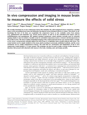
In vivo compression and imaging in mouse brain to measure the effects of solid stress
We recently developed an in vivo compression device that simulates the solid mechanical forces exerted by a growing tumor on the surrounding brain tissue and delineates the physical versus biological effects of a tumor. This device, to our knowledge the fi rst of its kind, can recapitulate the compressive forces on the cerebellar cortex from primary (e.g., glioblastoma) and metastatic (e.g., breast cancer) tumors, as well as on the cerebellum from tumors such as medulloblastoma and ependymoma. We adapted standard transparent cranial windows normally used for intravital imaging studies in mice to include a turnable screw for controlled compression (acute or chronic) and decompression of the cerebral cortex. The device enables longitudinal imaging of the compressed brain tissue over several weeks or months as the screw is progressively extended against the brain tissue to recapitulate tumor growth – induced solid stress. The cranial window can be simply installed on the mouse skull according to previously established methods, and the screw mechanism can be readily manufactured in-house. The total time for construction and implantation of the in vivo compressive cranial window is <1 h (per mouse). This technique can also be used to study a variety of other diseases or disorders that present with abnormal solid masses in the brain, including cysts and benign growths.

测试盒代表与眼部相关的脑损伤......
三种不同的脑神经调节运动。脑神经 III、IV 和 VI。脑神经 III 支配上直肌和下直肌,使瞳孔上下移动。脑神经 VI 支配外直肌,使瞳孔向外拉,然后脑神经 III 支配内直肌,使瞳孔向内拉。通过这种方式,人们可以通过观察是大运动受损还是协调受损来区分影响神经或通路的病变。
认知障碍和狼疮脑雾背后的故事
19 NPSLE manifestations Asceptic meningitis Cerebrovascular disease Demyelinating syndrome Headache Movement disorder Myelopathy Seizure disorders Acute confusional state Anxiety disorder Cognitive dysfunction Mood disorder Psychosis Acute inflammatory polyradiculoneuropathy Autonomic disorder Single/multiplex mononeuropathy Myasthenia gravis Cranial neuropathy Plexopathy多神经病
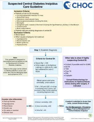
可疑的中央糖尿病临时护理指南
• s/p trans-sphenoidal resection of mass • Acute Brain Injury • Hypoxemic insult/vascular injury • Neurosurgical procedures involving the brain • Meningitis • Congenital cysts / masses of the brain involving the hypothalamus, pituitary or infundibulum • Cranial tumors • Brain malformations • Patients with pre-existing diagnosis of central DI Exclusion Criteria: • Renal failure • Other多尿的原因不排除:
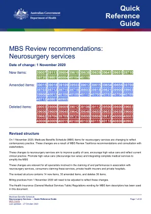

颅内突变病 - 神经手术
内镜下辅助条颅骨切除术(也称为内窥镜辅助毛切除术)是一种用于颅骨突变性手术的新方法。与传统方法一样,神经外科医生和整形外科医生删除了闭合的缝合线 - 但与传统方法不同,内窥镜手术不包括手术期间的颅骨重塑。这种微创手术通常是针对四个月以下的婴儿进行的,因为它取决于脑生长极快,以帮助重新定位颅骨。内窥镜辅助舒道切除术通常在手术室需要更少的时间,并且需要较短的住院时间。内窥镜手术进行颅骨突变病后,孩子将戴颅骨头盔以帮助重塑头骨。有关更多信息,请访问suturontromy.org
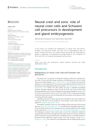
神经rest和儿子:神经rest细胞和雪旺细胞前体在发育和腺体胚胎发生中的作用
神经rest是一群多能迁移细胞的种群,在神经术期间从神经板的边界分离,并分化成成人生物体中各种器官的细胞(图1;表1)(他,1868年)。根据新的头部假设,正是神经波峰和表皮姿势的外观导致了弦脉的多样化和广泛分布(Gans and Northcutt,1983; Martik and Bronner,2021年)。神经rest细胞经历上皮 - 间质转变的阶段,并开始迁移到身体的远处。NCC与日益增长的神经以及转录特征的变化(SNAI1,SOX9/ 10,FOXD3,PAX3,PAX3和其他NCCS; SOX10,SOX2,SOX2,NRG1)的接触,SOX10,SOX2,NRG1,NRG1,NRG1),结果是Schwann细胞前体的形成,其发展依赖于AXT的迁移,并依赖于AXT的迁移。取决于其起源和定向迁移的位置,整个神经rest细胞(NCC)的种群可以分为几组:颅,树干,心脏和迷走NCCS(Achilleos and Trainor,2012年)。在哺乳动物中,颅NCC会产生颌骨和内耳的软骨和骨结构(Couly等,1998; Freyer等,2011)。此外,该细胞种群产生了牙齿的牙本质,额骨过程的骨骼以及颅神经的周围神经元和神经胶质(Leitevre,1978; Chai et al。,2000;Méndez-Maldez-Maldez-Maldonado et al。,2020)。颅内NCC还分化为The skeletogenic potential of the cranial neural crest has been extensively studied and documented from vertebrates, although cells of the trunk neural crest may also contribute to skeletal components in some animals like the contribution of NCCs to the differentiation of the plastron bones (abdominal carapace bones) of the turtle Trachemys scripta ( Cebra- Thomas et al., 2007 ).