XiaoMi-AI文件搜索系统
World File Search SystemPERCENTAGE
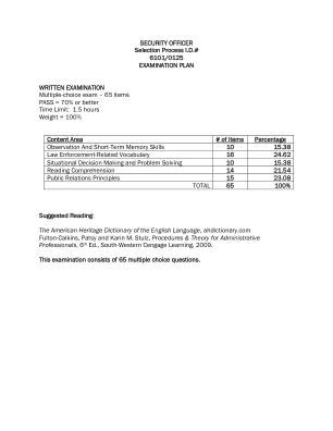
安全官
Content Area # of Items Percentage Observation And Short-Term Memory Skills 10 15.38 Law Enforcement-Related Vocabulary 16 24.62 Situational Decision Making and Problem Solving 10 15.38 Reading Comprehension 14 21.54 Public Relations Principles 15 23.08 TOTAL 65 100% Suggested Reading: The American Heritage Dictionary of the English Language , ahdictionary.com Fulton-Calkins, Patsy and Karin M. Stulz, Procedures &行政专业人员的理论,第6版,西南cengage学习,2009年。此检查由65个多项选择问题组成。
重新列出了dna de dna delinfócitost
Figura 1: Desenho esquemático dos componentes presentes nas redes extracelulares de DNA em neutrófilos ................................................................................................................................................17 Figura 2: Representação esquemática da via da Netose ........................................................................19 Figura 3: Desenho experimental .................................................................................................................28 Figura 4: Estratégia de análise para avaliar a frequência das populações de CD14+,CD19+, CD4+ e CD8+ após a Marcaçãocomanticorpos antisubpopulaçõescelulares eaquisiçãoporcitometriade fluxo。..................................................................................................................................................................................................................................................................................................................................................................................................................................................................................................................................................... 38图5:富集淋巴细胞的细胞组成的细胞仪分析。。...................................................................................................................................................39 Figura 6: Avaliação da porcentagem de morte celular por citometria de fluxo ................................... 40 Figure 7: Evaluation, in lymphocyte -enriched crops, by confocal microscopy, of the occurrence of extracellular DNA. 在 ................. 51 Figure 15: SME evaluation of CD4+ and CD8+ T lymphocytes................................... 40 Figure 7: Evaluation, in lymphocyte -enriched crops, by confocal microscopy, of the occurrence of extracellular DNA.在................. 51 Figure 15: SME evaluation of CD4+ and CD8+ T lymphocytes.................. 51 Figure 15: SME evaluation of CD4+ and CD8+ T lymphocytes........................................................................................................................................................................................................................................................................................................................................................................................................................................................................................................................ .......................................................................................................................................................................................................................................................................................................................................................................................................................................................................................................................................................................... 46 Figure 11: Extracellular networks blushed by DAPI do not have actin. ............................... 47 Figure 12 Electronic microscopy assessment of scanning the presence of extracellular networks in lymphocyte -enriched crops. .......................................................................... 50 Figure 13 Electronic Microscopy Evaluation of Internal Morphology Transmission of Lymphocytes. ............................................................................................................................................................................................................................................................................................................................................................................................................................................................................................................................................................................... ............................................................................................................................................................................................................................................................................................................................................................................................................................................................................................................................................................................... ......................................................... 52 Figure 16: Evaluation of the percentage of cell death by CD4+ and CD8+ T lymphocyte flow cytometry. .............................................................................................................................................................................................................................................................................................................................................................................................................................................................. 54 在........................................... 46 Figure 11: Extracellular networks blushed by DAPI do not have actin................................ 47 Figure 12 Electronic microscopy assessment of scanning the presence of extracellular networks in lymphocyte -enriched crops. .......................................................................... 50 Figure 13 Electronic Microscopy Evaluation of Internal Morphology Transmission of Lymphocytes. ............................................................................................................................................................................................................................................................................................................................................................................................................................................................................................................................................................................... ............................................................................................................................................................................................................................................................................................................................................................................................................................................................................................................................................................................... ......................................................... 52 Figure 16: Evaluation of the percentage of cell death by CD4+ and CD8+ T lymphocyte flow cytometry. .............................................................................................................................................................................................................................................................................................................................................................................................................................................................. 54 在............................... 47 Figure 12 Electronic microscopy assessment of scanning the presence of extracellular networks in lymphocyte -enriched crops........................................................................... 50 Figure 13 Electronic Microscopy Evaluation of Internal Morphology Transmission of Lymphocytes. ............................................................................................................................................................................................................................................................................................................................................................................................................................................................................................................................................................................... ............................................................................................................................................................................................................................................................................................................................................................................................................................................................................................................................................................................... ......................................................... 52 Figure 16: Evaluation of the percentage of cell death by CD4+ and CD8+ T lymphocyte flow cytometry. .............................................................................................................................................................................................................................................................................................................................................................................................................................................................. 54 在.......................................................................... 50 Figure 13 Electronic Microscopy Evaluation of Internal Morphology Transmission of Lymphocytes................................................................................................................................................................................................................................................................................................................................................................................................................................................................................................................................................................................ ........................................................................................................................................................................................................................................................................................................................................................................................................................................................................................................................................................................................................................................ 52 Figure 16: Evaluation of the percentage of cell death by CD4+ and CD8+ T lymphocyte flow cytometry. .............................................................................................................................................................................................................................................................................................................................................................................................................................................................. 54 在......................................................... 52 Figure 16: Evaluation of the percentage of cell death by CD4+ and CD8+ T lymphocyte flow cytometry............................................................................................................................................................................................................................................................................................................................................................................................................................................................... 54 在.............................................................................................................................................................................................................................................................................................................................................................................................................................................................. 54在.............................................. 55 Figure 18 Evaluation by CD107 + cell frequency flow cytometry 56 Figure 19: Immunofluorescence of CD8 +, Lets and CD107. ........................ 58 Figure 21: Evaluation of cellular composition of patient injuries through histopathological analysis by hematoxylin/eosin coloration. ........................................................................59 Figura 22: Avaliação da composição celular de lesões de pacientes com LTA ..................................60 Figura 23 : Avaliação por microscopia confocal de ETs em lesões de pacientes da forma cutânea da LTA . ............................................................................................................................................................................................................................................................................................................................................................................................................................................................................................................................................................................. ............................................................................................................................................................................................................................................................................................................................................................................................................................................................................................................................................................................. .............................................................................................. 65.............................................. 55 Figure 18 Evaluation by CD107 + cell frequency flow cytometry 56 Figure 19: Immunofluorescence of CD8 +, Lets and CD107......................... 58 Figure 21: Evaluation of cellular composition of patient injuries through histopathological analysis by hematoxylin/eosin coloration. ........................................................................59 Figura 22: Avaliação da composição celular de lesões de pacientes com LTA ..................................60 Figura 23 : Avaliação por microscopia confocal de ETs em lesões de pacientes da forma cutânea da LTA . ............................................................................................................................................................................................................................................................................................................................................................................................................................................................................................................................................................................. ............................................................................................................................................................................................................................................................................................................................................................................................................................................................................................................................................................................. .............................................................................................. 65........................ 58 Figure 21: Evaluation of cellular composition of patient injuries through histopathological analysis by hematoxylin/eosin coloration.........................................................................59 Figura 22: Avaliação da composição celular de lesões de pacientes com LTA ..................................60 Figura 23 : Avaliação por microscopia confocal de ETs em lesões de pacientes da forma cutânea da LTA .............................................................................................................................................................................................................................................................................................................................................................................................................................................................................................................................................................................. ........................................................................................................................................................................................................................................................................................................................................................................................................................................................................................................................................................................................................................................................................... 65.............................................................................................. 65

WISETECH全球性别薪酬差距报告2025
报告期间的付款被排除在外。正百分比表明男性的薪水平均比女性的收入高或中位数。A negative percentage indicates women are paid more than men on average or at the median 11 Prior to 2023-24, WGEA's GPG calculation excluded CEO, Heads of Business (HOB), overseas managers (OSM), and casual managers (CM) 12 WGEA's GPG calculation for 2023-24 includes CEO, HOB and CM (where submitted) and continues to exclude OSM 13 Total remuneration includes base薪水,退休金,薪酬权益,加班,奖金和其他额外付款。平均总报酬为最接近的$ 1,000

经济更新 - 2023年11月
- 2023年10月的年度通货膨胀率最大的贡献来自餐馆和咖啡馆(0.40个百分点);水果(0.35个百分点);个人护理设备,物品和产品(0.25个百分点);电力(0.24个百分点);乳制品(0.22个百分点);门诊护理服务(0.20个百分点);其他食品(0.18个百分点);蔬菜(0.18个百分点);海上的客运运输(0.17个百分点);和移动通信服务(-0.80个百分点)。

关于绿色供应链问题和收益的研究...
ABSTRACT (i) LIST OF TABLES (ii) LIST OF CHARTS (iii) 1 INTRODUCTION 1.1 Introduction 1 1.2 Industry profile 8 1.3 Company profile 12 1.4 Statement of the problem 17 1.5 Needs of the Study 17 1.6 Scope of the Study 18 1.7 Objective of the study 18 2 REVIEW OF LITERATURE 2.1 Review of Literature 20 3 RESEARCH METHODOLOGY 3.1 Research Design 36 3.2 Sample size 37 3.3 Source of Data 37 3.4 Tools of Analysis 38 4 DATA ANALYSIS AND INTERPRETATION 4.1 Percentage分析41 4.2卡方64 4.3相关66 5调查结果,建议和结论5.1研究结果68 5.2研究建议69 5.3研究限制70 5.4结论70参考附件 - 问卷
冷凝财务报告 - 补充数据
核心营业收入为38亿美元( +5%, +13%CC),主要由较高的净销售额驱动,部分地被SG&A和R&D投资较高。美元的核心营业收入增长受阿根廷货币贬值的影响负2个百分点的影响。核心营业收入利润率为净销售额的33.5%,下降了1.0个百分点(+1.0百分点CC)。其他收入占销售额的百分比增加了0.1个百分点(CC)。销售商品的核心成本占销售额的百分比,下降了0.2个百分点(CC)。核心研发费用占净销售额的百分比,减少了1.1个百分点(CC)。核心SG&A支出额的净销售额的百分比下降了0.1个百分点(CC)。核心其他收入和净销售百分比的费用降低了0.5个百分点(CC)。

科学和社会科学中学课程的气候变化和可持续性
图1。根据环境,可持续性和气候变化的重点,科学和社会科学课程的百分比。 图2。 内容范围在环境,可持续性和气候变化上(标准化参考)。 图3。 关注环境,可持续性和气候变化内容的百分比。 图4。 比较NCFS和科学与社会科学课程(平均标准化参考文献)。 图5。 内容变化的内容百分比。 图6。 按国家和地区划分气候变化的内容百分比。 图7。 在环境上的内容百分比。 图8。 按国家和地区划分的环境中内容的百分比。 图9。 可持续性内容的百分比。 图10。 按国家和地区划分可持续性的内容的百分比。 图11。 科学和社会科学课程的百分比,按关注环境的重点,可持续性根据环境,可持续性和气候变化的重点,科学和社会科学课程的百分比。图2。内容范围在环境,可持续性和气候变化上(标准化参考)。图3。关注环境,可持续性和气候变化内容的百分比。图4。比较NCFS和科学与社会科学课程(平均标准化参考文献)。图5。内容变化的内容百分比。图6。按国家和地区划分气候变化的内容百分比。图7。在环境上的内容百分比。图8。按国家和地区划分的环境中内容的百分比。图9。可持续性内容的百分比。图10。按国家和地区划分可持续性的内容的百分比。图11。科学和社会科学课程的百分比,按关注环境的重点,可持续性

公共咨询报告
Suburb Participants Percentage HYDE PARK, QLD 1 0.40% IDALIA, QLD 6 2.43% JENSEN, QLD 4 1.62% KELSO, QLD 7 2.84% KIRWAN, QLD 20 8.13% MAGNETIC ISLAND, QLD 2 0.81% MOUNT LOUISA, QLD 7 2.84% MOUNT LOW, QLD 2 0.81% MUNDINGBURRA, QLD 7 2.84% MYSTERTON, QLD 1 0.40%Nelly Bay,QLD 4 1.62%北区,QLD 12 4.87%Oak Valley,QLD 1 0.40%Oonoonba,QLD 5 2.03%野餐湾,QLD 1 0.40%Pimlico,QLD 7 2.84 2.84%2.84%铁路,QLD 5 2.03%Rasmussen,Qld Rasmussen,Qld 1 0.40%0.40%,QLD 3 1. 3.65%Thingowa Central BC,QLD 1 0.40%Toomulla,QLD 1 0.40%汤斯维尔城,QLD 13 5.28%Vincent,QLD,QLD 4 1.62%西区,QLD 15 6.09%Wulguru,QLD 4 1.62%
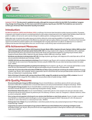
AFIB质量度量
• Angiotensin-Converting-Enzyme Inhibitors (ACEI)/Angiotensin Receptor Blocker (ARB) or Angiotensin Receptor-Neprilysin Inhibitor (ARNI) prescribed prior to discharge (when Left ventricular ejection fraction (LVEF) <40): Percentage of patients with a diagnosis of atrial fibrillation (AF) or atrial flutter with heart failure (HF) with左心室射血分数LVEF≤40,他们被处方了血管紧张素转换 - 酶酶抑制剂(ACEI)/血管紧张素受体阻滞剂(ARB)或血管紧张素受体抑制剂 - 涅台环蛋白抑制剂(ARNI)。AHAAF1•在出院前开处方的β受体阻滞剂(当LVEF≤40):年龄≥18岁的患者百分比,诊断为AF或心房颤动,其LVEF≤40在出院前被处方为Beta阻滞剂。ahaaf2。•出院前记录的CHA2DS2-VASC风险评分:年龄≥18岁的患者百分比,具有非瓣膜和生物假体瓣膜房颤或心房颤音,用于使用CHA2DS2-VASC风险评分标准对其进行评估,以评估其医疗记录。ahaaf3。•FDA批准的批准的抗凝治疗:出院前处方:患者百分比≥18岁,没有瓣膜性房颤或心房颤动,这些抗凝或心房的扑动,这些抗凝被开处方华法林或其他FDA批准的抗凝药物,以预防血栓栓塞。AHAAF5•AF患者的冠状动脉疾病患者(CAD),CVATYPE/TIA,周围血管疾病(PVD)或糖尿病患者的分泌:CAD,CAD型/TIA,TIA,PVD或糖尿病患者百分比。ahaaf6AHAAF4 • Prothrombin Time/International Normalized Ratio (PT/INR) Planned follow-Up documented prior to discharge for warfarin treatment: Percentage of patients, age ≥18 years, with nonvalvular, valvular or bioprosthetic valve AF or atrial flutter who have been prescribed warfarin and who have a PT/INR follow-up scheduled prior to hospital discharge.
2019 年底英国 GDP 持平 - 大伦敦政府
除了产出,伦敦的劳动力市场状况也保持了弹性,英国国家统计局的数据显示,截至 12 月的三个月内,伦敦 16-64 岁就业率为 75.5%,比上一季度上升 0.9 个百分点,比去年同期上升 0.7 个百分点。整个英国的就业率比去年同期上升了 0.6 个百分点,达到 76.5%。与此同时,伦敦的失业率(失业人数占劳动力的百分比)接近历史最低水平,为 4.3%,比上一季度下降 0.3 个百分点,比去年同期下降 0.2 个百分点。英国失业率估计为 3.8%,比上季度下降 0.1 个百分点,比去年同期下降 0.2 个百分点。